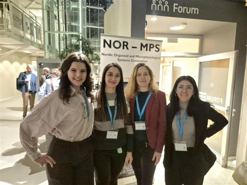
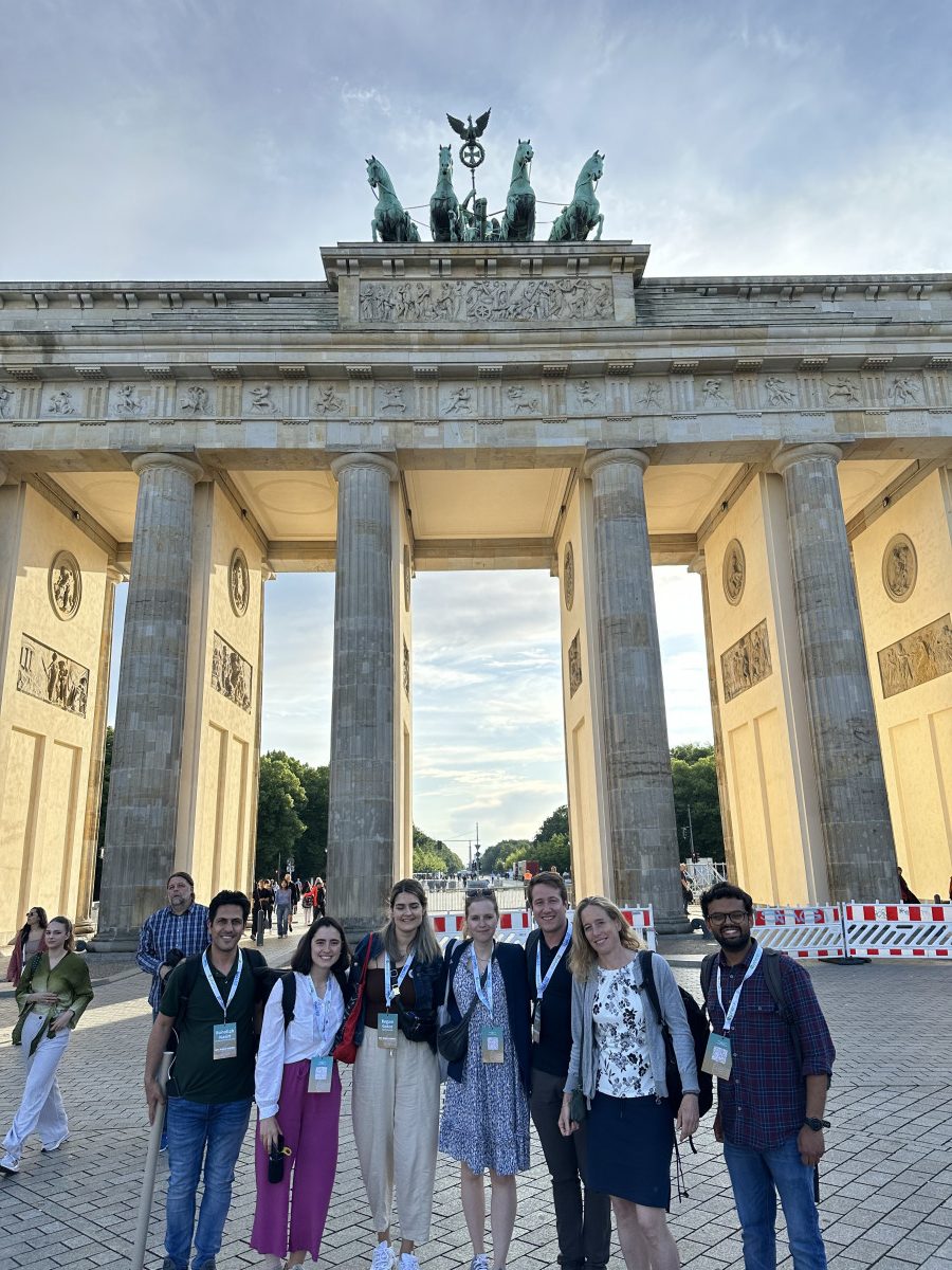
Our group picture at the NOR-MPS 2026 venue. From left to right: Katia, Ana, Anna, and Angela.
Herland Lab participated in the Nordic Organoid and Microphysiological Systems Convention (NOR-MPS) 2026, held at Forskningsparken, Oslo Science Park. The one-day symposium brought together academic and industry researchers across the Nordic region to share advances in organoids, microphysiological systems, and organ-on-chip technologies.
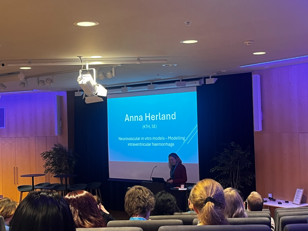
Anna presenting on in vitro intraventricular haemorrhage model.
From Herland Lab, participants included Angela (PhD student), Ana (Postdoc), and Katia (MSc student). Anna delivered an oral presentation on ‘Neurovascular in vitro models for modelling intraventricular haemorrhage‘, highlighting our lab’s work on advanced human-relevant platforms for studying brain injury mechanisms. Meanwhile, Ana was awarded 2nd place for Best Poster Presentation.
Ana was awarded 2nd place for Best Poster Presentation.
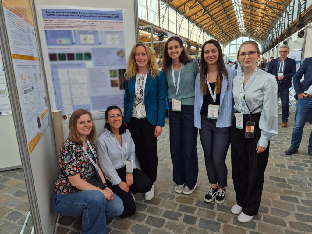
Herland Lab participated in the Microphysiological System (MPS) World Summit 2025 in conjunction with theEUROoCS Annual Meeting 2025 in Brussels, Belgium on the 9th to 13th June 2025.
Our group picture in front of one of the posters presented at MPS/EUROoCS 2025. From left to right: Julia, Angela, Anna, Ludovica, Ana, and Justina.
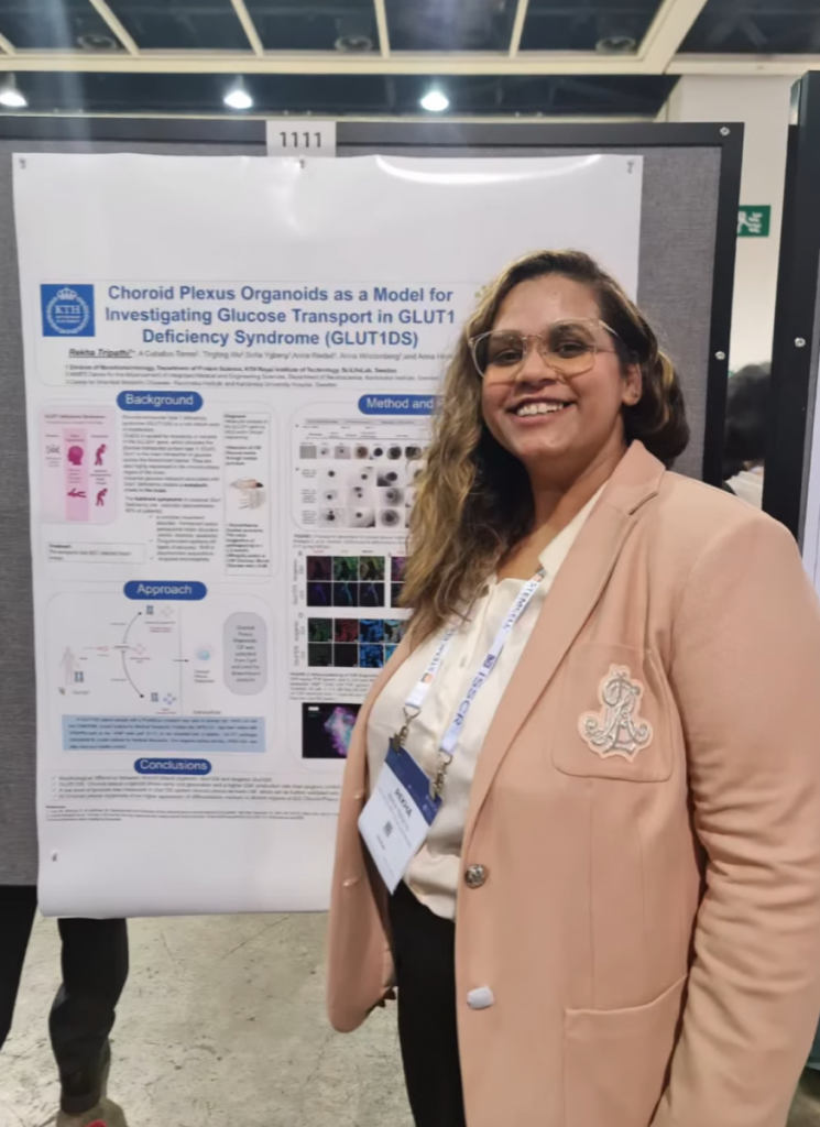
We also have a poster presented at the International Society for Stem Cell Research (ISSCR) Annual Meeting 2025 in Hong Kong from the 11th to 14th June 2025.
As the spring semester comes to a close, we are taking a moment to reflect on the many milestones our group has celebrated. May has been a month of transformation at Herland Lab, and we are proud to celebrate the brilliant minds who have recently completed important chapters in their journeys with us! From defending a PhD to wrapping up MSc research projects that pushed boundaries across disciplines, here is our story.
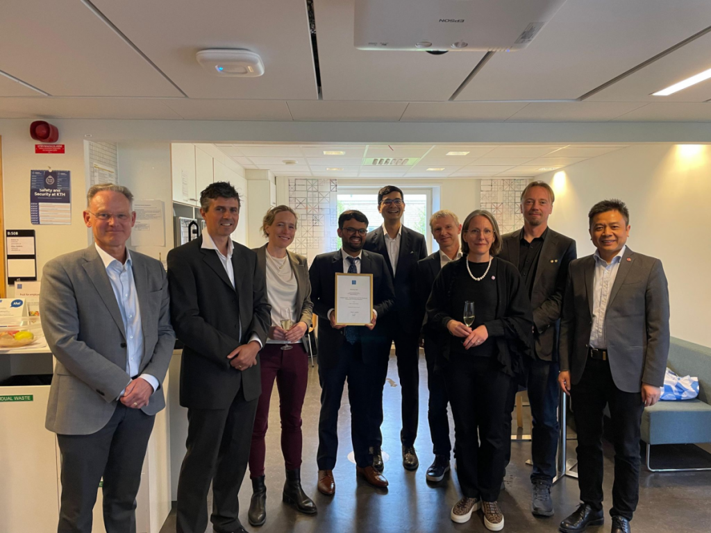
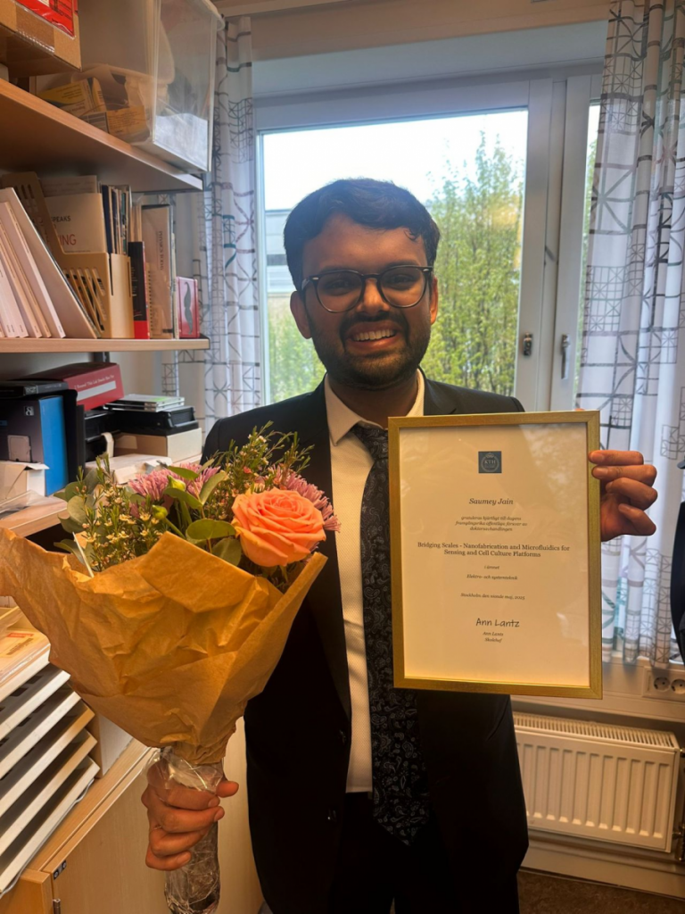
PhD Defense: Bridging Scales in Biosensing and Cell Culture
On May 9th, our PhD student Saumey Jain successfully defended his thesis: “Bridging Scales – Nanofabrication and Microfluidics for Sensing and Cell Culture Platforms“. His research journey explored the intersection of physics, engineering, and biology; from the development of nanoscale biosensing platforms such as nanogaps and solid-state nanopores, to applying microfabrication techniques for improving iPSC-derived neuron differentiation.
He now brings that scientific depth into the innovation pipeline as he joins HØIBERG (European Patent Attorneys), helping other researchers bring their technologies to life. Good luck, Saumey, and thank you for being the backbone of our lab!
MSc Research Highlights: Bridging Disciplines
We also want to give a standing ovation to our MSc students, who just wrapped up their projects with impressive progress and dedication, both in the lab and during their final presentations! Each project reflects the interdisciplinary spirit we strive for. Here are just a few of the exciting directions they pursued:
Gabriel Gyllensting (Karolinska Institutet, Medical Program): Organ-on-Chip Model for BBB Invasion of E. coli in Bacterial Meningitis
Shaishav Shah (Stockholm University, MSc Neurochemistry with Molecular Neurobiology): Modeling the Choroid Plexus using 2D and 3D In Vitro Models
Jacqueline Clark (Stockholm University, MSc Analytical Chemistry): Real Time Continuous Monitoring of Neuromuscular Chips using Electrochemical Biosensors
Liisa Loel (KTH, MSc Molecular Techniques in Life Science): Evaluation of iPSC-derived Neuromuscular Cells in a Novel Neuroenergetics-on-chip (NEoC) Model
Danhong Song (Karolinska Institutet, MSc Translational Physiology and Pharmacology): 3D Printed Bioscaffolds for Macrophage Modulation and Preserved Insulin Secretion in Pancreatic Islet Transplantation
Nadja Widén (KTH, MSc Engineering Physics): Comprehensive Study of Drug-loaded Bioscaffolds
International Visits and Exchanges
This year, we have been lucky to both host and send out researchers across borders, reflecting our commitment to cross-cultural science, shared learning, and building bridges across research ecosystems.
Earlier this semester, we had the pleasure of hosting Thanawin Jantheang and Ludovica Montesi, two visiting PhD students who enriched our lab with their work on brain organoids and the NEoC model.
Meanwhile, one of our PhD students, Meike Bleeksma, is currently spending a year at the University of Tokyo with Prof. Shoji Takeuchi’s lab, diving into new collaborations and techniques. Another PhD student, Yunfan Lin, will also soon begin his one-year exchange at Shanghai Jiao Tong University (SJTU) next semester.
Member Transitions: Farewells and New Faces
This spring also marked a bittersweet moment as we said goodbye to one of our postdocs, Sebastian Buchmann, who is now continuing his journey also at HØIBERG. At the same time, we are thrilled to welcome two new postdocs into the group, Ana Spencer and Eleni Mitoudi-Vagourdi, to strengthen our core research areas and spark new directions. We are also excited to celebrate Angela Ceballos, who has taken her first step into the PhD program at Karolinska Institutet; congratulations!
To everyone who has defended, transitioned, presented, traveled, or joined us this semester — thank you! Your energy, curiosity, and contributions make Herland Lab more than just a place of science; they make it a growing, thriving community. Here’s to the next chapter! 🎉
We are proud to share that our group leader, Anna Herland, has been appointed full professor in Hybrid Bioelectric Systems at KTH during the 2024 Professorial Installation Ceremony! KTH highlighted Professor Herland’s research in a recent video feature, which can be checked out here: [Link to KTH website]
Professor Herland’s research focuses on the development of Organ-on-Chip (OoC) systems, microphysiological platforms that replicate human tissue environments using patient-specific hiPSC-derived cells. These systems are integrated with organic bioelectronics to enable real-time functional assessments and dynamic readouts of tissue responses.
Her vision has shaped the foundation of our lab’s interdisciplinary approach, combining stem cell engineering, microfluidics, and organic electronics to explore how the brain and body interact at the cellular level. A major area of our work is building advanced models of the human neurovascular unit, where we study neuronal activity, metabolic dynamics, and blood-brain barrier function in health and disease. These systems are powerful tools laying the groundwork for more predictive drug testing and new tools in personalised medicine.
The group gathered to celebrate this milestone with a small lab event, complete with professor sashes and celebratory crowns!
One of our collaborative works with Karolinska University Hospital was featured in an interview by KTH, with the theme of ‘3D Printing and Polymer Chemistry Revolutionize Diabetes Treatment‘. This project has been supported by the Health, Medicine, and Technology (HMT) initiative from KTH and Region Stockholm.
Together with Prof. Lisa Juntti-Berggren from the Signal Transduction research group at KI, we are developing a novel method to improve pancreatic islet transplantation into the anterior chamber of the eye (ACE). Our doctoral student, Jessika Jessika, is currently leading the exploratory phase to address the challenges in manufacturing drug-releasing microstructure along with some of the MSc students in the group.
In our latest research, we developed an OECT-based biosensor with a p-type polymer bithiophene–thienothiophene copolymer with tetraethylene glycol side chains, p(g42T-TT), as the channel material. The xanthine biosensor aims to monitor the quality of meat and fish products, providing freshness predictions with respect to pre-defined expiration dates. The biosensor detects the xanthine level, which reflects the degradation of meat and fish products. The results show the time-dependent xanthine in fish samples from day 0 to day 6.
This initiative, funded by the Eureka Eurostars program, aims to develop a cutting-edge Gut-Brain-Axis (GBA) on-chip platform integrating advanced organ-on-chip technologies, including in vitro blood-brain barriers and co-cultured anaerobic microbiota. Next-generation hardware for automated culturing, monitoring, and analysis will also be investigated to address critical gaps in current drug discovery methods. This platform will provide new insights into drug absorption, blood-brain barrier permeability, and drug toxicity evaluation to enhance our understanding of the Gut-Brain-Axis, with the potential to revolutionise therapeutic development for neurological and mental health conditions.
Stay tuned for more updates on the progress of this exciting research!
In our latest research, we present the new conjugated polymer p(g42T-T)-8% OH, a promising material for simplifying the interface between biological systems and electronics. This polymer contains hydroxyl groups, enabling straightforward chemical modifications to control cell adhesion and growth on its surface. We demonstrate the fabrication of organic electrochemical transistors (OECTs) using p(g42T-T)-8% OH, which were successfully employed to monitor the formation of cell barriers in vitro. This research provides a valuable tool for studying biological systems and offers a pathway for developing customizable bioelectronic devices that more effectively interface with biological tissues.
The first article introduces feedback-controlled electromigration to fabricate stable gold nanogap tunnel junctions for single-molecule sensors capable of operating robustly across various liquid and gaseous environments. We systematically examine both the electrical behaviour and the yield of junction formation across different media, with attention to how specific operating conditions influence stability and performance. The findings not only advance our understanding of tunnel junction scalability and production but also enhance the potential for integrating these sensors into practical, compact devices.
In the second article, we describe an innovative measurement setup that supports high-bandwidth (>10 kHz) and low-current (pA–nA) measurements, crucial for the effective probing of these tunnel junctions. By integrating a custom two-terminal probe with a 100 kHz bandwidth amplifier and automating the data acquisition within a noise-reducing Faraday cage, we have streamlined the scalability and improved the precision of these measurements.
Blending polystyrene with n-type conjugated polymer p(N-T) is demonstrated to produce high-performing OECT with drastically reduced conjugated polymer required to achieve functionality. By using commodity polymers such as polystyrene, the organic semiconductors’ material consumption could then be used sparsely, leading to the cost-effective and sustainable development of organic transistors.
The established protocols aim to be a sustainable alternative to perform device fabrication with reduced dependency on cleanroom facilities, which involve a significant amount of solvents and chemical developer baths in the process. The work also demonstrated the possibility of performing rapid prototyping with micropatterning based on ultrafast laser technology to manufacture different types of organic bioelectronics devices.
Understanding the mechanism behind CNS energy metabolism and their role on brain function is crucial to answer the relevant questions in human neurological conditions. Establishing a physiologically relevant in vitro models of neuroenergetics requires comprehensive understanding of different cell types processes and interaction, also various metabolic pathways and regulatory mechanisms. The article highlights important keypoints in the development of human in vitro model of neuroenergetics, covering both the biology and technology perspectives.
The work has successfully demonstrated the first microfluidics platform to redirect iPSCs toward becoming neural stem cells. Here, a simple and cost-efficient microfluidic platform is developed to reprogram human fibroblasts into induced pluripotent stem cells (iPSCs) and further differentiate them into neural stem cells with boosted neural stem cell generation commitment. With further customization, the platform could be adapted for differentiation into other cell types, marking an important step towards making personalized cell-based therapies for Alzheimer’s and Parkinson’s disease more accessible.
Microphysiological systems, like organ-on-a-chip models, show promise for healthcare and drug development. Clear standards are needed for their effective use. Collaborative efforts are underway to establish these standards, with input from regulators, academia, and industry. This framework aims to foster discussion and enhance the impact of microphysiological systems.
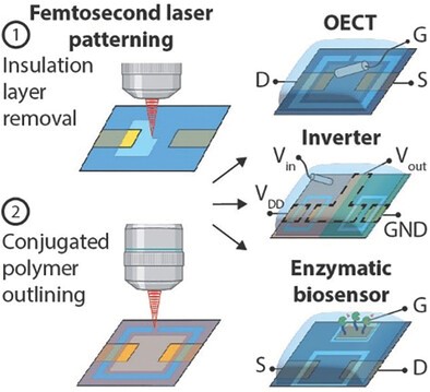
By employing femtosecond laser technology, we achieved high-resolution patterning of insulating and (semi)conducting polymers with a resolution down to 2 micrometers, providing a cost-effective alternative to traditional cleanroom-based microfabrication methods. This versatile and scalable approach is particularly well-suited for prototyping, therefore accelerating OECT research and facilitating their widespread use in applications such as biosensors or logic gates. Our work marks a significant step towards more accessible and efficient OECT fabrication methods, offering potential benefits for a variety of advanced organic bioelectronic applications.
This study introduces a pioneering approach for transplanting biohybrid microstructures into the anterior chamber of the eye. This technique combines biological cells with sensors to detect subtle physiological responses with precision. Overcoming challenges related to pupillary dynamics, the method ensures secure transplantation and prolonged functionality of microstructures housing pancreatic islets. Even more striking, these islets develop a blood supply, offering exciting prospects for improved disease modeling, treatment efficacy, and engineered tissue vascularization. This breakthrough redefines possibilities in bioengineering and regenerative medicine, providing hope for enhanced medical treatments.
The Herland Lab spent an intense and enjoyable week in Berlin, Germany at Microphysiological Society World Summit which was held together with the European Organ-on-Chip conference.
Sebastian gave a talk in the session on “Microfabrication, Instrumentation & Sensors” and Laura presented the work done as a part of master thesis in the session on “MPS for Vascularization”. Julia, Rohollah, Saumey, and Begum presented posters with their latest research results.
Julia, Begum, Sebastian and Laura were also awarded travel grants.
In this work, we have developed a 3D-printed platform that enables precise control and in-depth study of the interaction between astrocytes and neurons. To achieve this, we utilized a commercially available printing resin IP-Visio (Nanoscribe), with non-autofluorescent and non-cytotoxic properties. We successfully printed a neurite guidance platform, which served as a guiding structure for the outgrowth of neurites from neurons. By employing a probabilistic two-step cell seeding approach, we were then able to control the interaction points between neurons and astrocytes. This innovative approach simplifies the modeling of complex brain interactions, thereby facilitating further advancements in biomedical research and enhancing our understanding of the human brain.
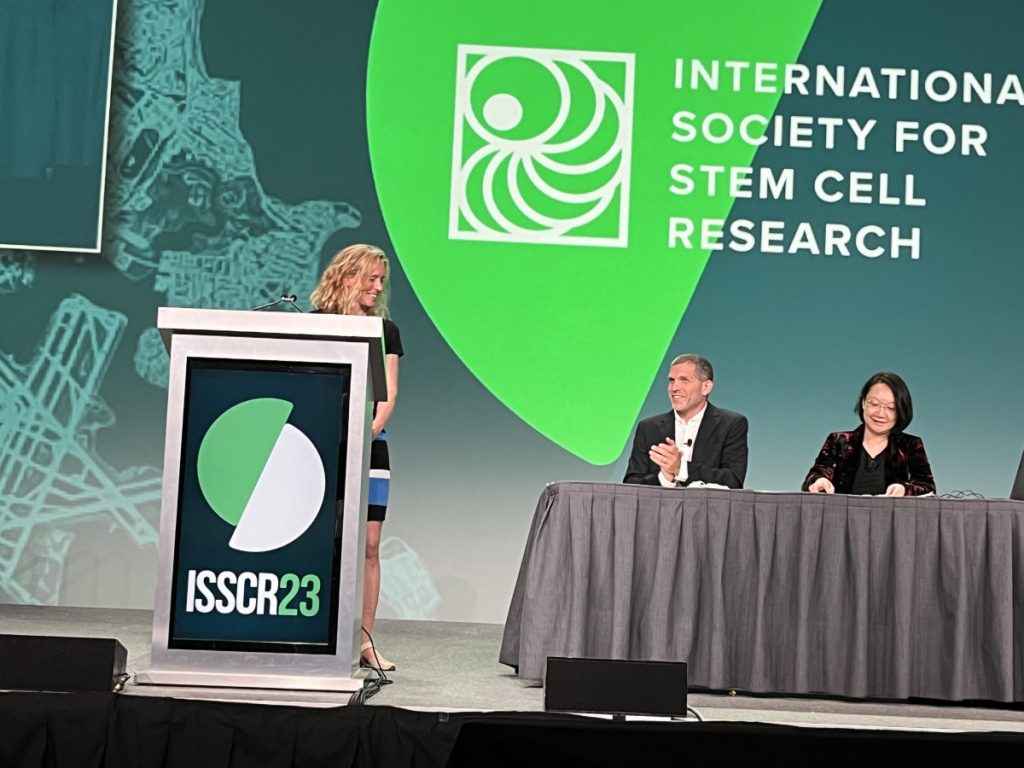
Prof. Anna Herland gave the plenary talk at The International Society for Stem Cell Research‘s Annual Meeting 2023, 14-17th June 2023 in Boston on next-generation in-vitro models for modeling development and disease.
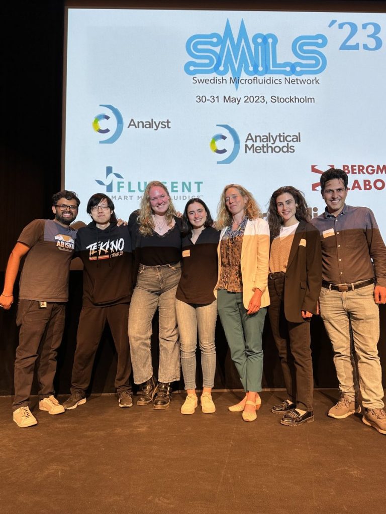
Herland Lab participated at the Swedish Microfluidics in Life Science conference in Stockholm on the 30th and 31st of May. Laura and Rohollah had oral presentations, whereas Yunfan, Monika, and Salem presented their research as posters. It was a great opportunity to network with the microfluidics network in Sweden as well as the neighboring countries.
Laura Benito-Zarza pitched her master thesis work today during the Nordic Organ-on-Chip networking event and won one of the best pitch awards, consisting of a travel grant in the Nordics to expand her network and experience in the Organ-on-Chip field. She is supervised by Dr. Julia Rogal and Dr. Alessandro Enrico and working to develop a hiPSC-derived microvascularized tissue model to recapitulate in vivo complexity for the study and treatment of complex diseases, such as Parkinson’s and Alzheimer’s.
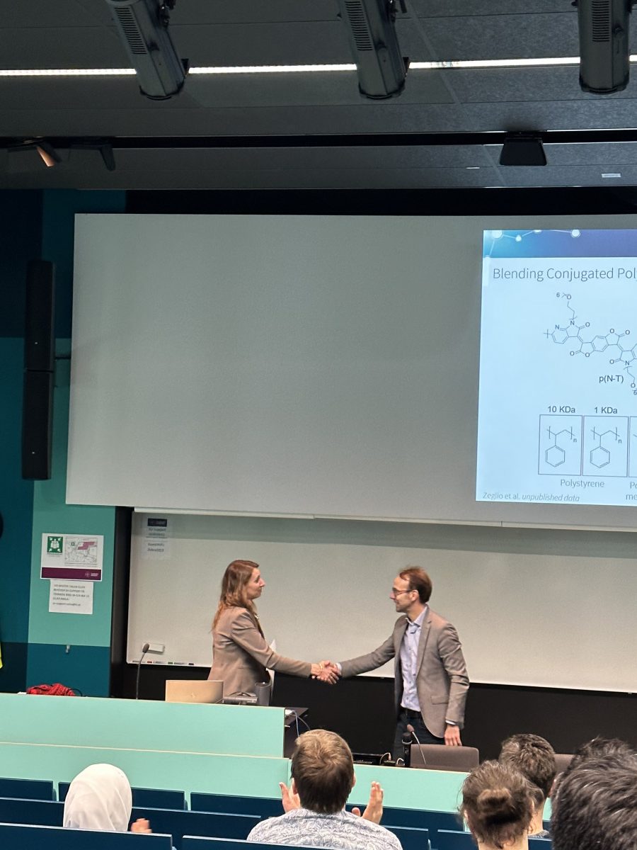
Dr. Erica Zeglio was admitted as a Docent in Bioelectronic Materials at KTH. She gave the docent lecture on ‘Organic bioelectronics: from sustainable materials chemistry to the interface with biology.’ The docentship in the Swedish system means she can supervise and be an opponent for a PhD candidate and is not supposed to lose badly.
This work highlights the development of peptide functionalized hyaluronan hydrogels for bioprinting. The study utilized neuroblastoma (SH-SY5Y) and glioblastoma (U87) cell lines and human fetal primary astrocytes (FPA) with a modular hyaluronan-based hydrogel system. It was observed that FPA had a higher degree of interaction with the hyaluronan-based gels compared to the cell lines. These engineered hydrogels enable the possibilities to bioprint, culture and maintain FPA and can thus facilitate the development of more elaborate neural and astrocytic tissue and disease models.
Prof. Anna Herland and Dr. Erica Zeglio are awarded grants from the Swedish Research Council (Vetenskaprådet).
Refining neurovascular in vitro models (Prof. Anna Herland, co PIs: Ryan Hicks (AstraZeneca), Xenia Nikolakopoulou (KI), Jane Synnergren (University of Skövde))
All new drug candidates need to be evaluated for central nervous system (brain) penetration and sideeffects. Unfortunately, and especially for new drug types, the testing methods do not show how the drugs will work in humans. Here, we focus on developing and validating human functional Organ-on-Chip models of the brain and its vasculature. In this work, which is a collaboration with AstraZeneca around the theme to reduce and refine animal experiments, we especially focus on human specific drugs including viral vectors and protein or peptide-based drugs.
2. 2D and 3D in vitro models with organic electronic interfaces to electrogenic cells (PI: Prof. Anna Herland, co PIs: Prof. Max Hamedi (KTH), Prof. Frank Nikalus (KTH) and Dr. Erica Zeglio (KTH))
Electrodes are needed to stimulate and measure on electrically active tissues, such as the heart and brain. Electrodes are specifically used in electrophysiology, the studies of electrically active cells outside the body. This is an essential method in drug development and toxicity studies.
The main goal in this project is to create new methods to carry out electrophysiology in 3D and microfluidic cell culture, so called Organ-on-Chips. We will combine the development of new polymer materials, new transistor designs, and new fabrication methods for electroactivity measurements in 2D and 3D systems.
3. Biodegradable electronic polymers: from device components to in vivo monitoring technologies (VR Starting Grant, Dr. Erica Zeglio)
Temporary implants are currently based on electronics that dissolve in contact with water, limiting their application in contact with (water-based) biological fluids. With the project “Biodegradable electronic polymers: from device components to in vivo monitoring technologies”, the Team led by Dr. Zeglio will investigate device components that are stable in contact with water and degrade into nontoxic products by the action of living cells.
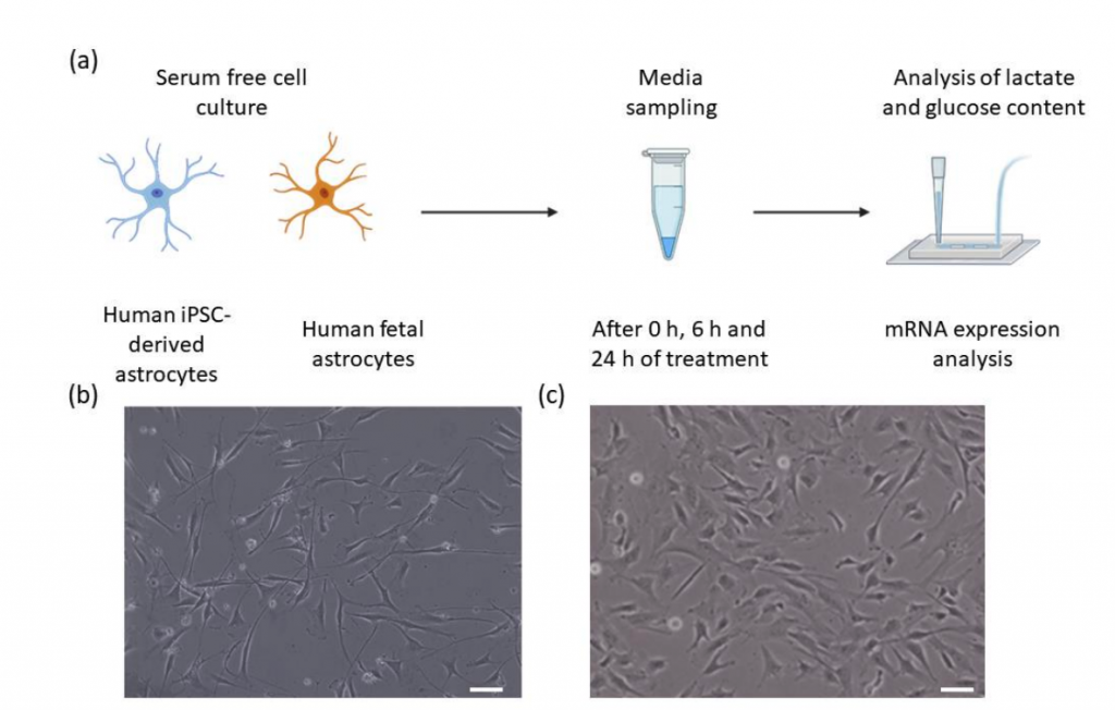
Astrocytes are one of the key cell types in brain for energy metabolism. In this research article, the metabolic parameters of astrocytes derived from two common sources including induced pluripotent stem cells (hiPSCs) and human fetal primary astrocytes (HFAs) were studied in a defined media. The glucose uptake and lactate production in astrocytes derived from hiPSC to HFA were compared using a flow-through biosensor. The study concluded that hiPSC-derived astrocytes are as glycogenic as their fetal counterparts, but their normalized metabolic turnover is lower.
Herland Lab (re)welcomes Kim and Yunfan after completing their Masters! Kim Roekevisch is back as a Research Engineer at KI, working on the targeting of the CNS using AAVs with Xenia and Julia. Yunfan Lin is also back as a Research Engineer at KTH, working on the development of biodegradable electrochemical sensors for food monitoring with Erica and Anna.
A long-standing question in tissue engineering is how to vascularize tissue models. This study made a breakthrough with a technology that can enable much more physiological vascularization. The article reports a novel patterning approach for collagen hydrogels, referred to as “cavitation molding”, to create 3D cavities that can be used as a template to form the microvasculature. This method enables the fabrication of relevant models to study complex tissue, such as tumors and neural tissue.
Vascularized cancer-on-a-chip model developed using cavitation molding of collagen
Dr. Julia Rogal has joined the Herland Lab as a new postdoctoral scholar at the beginning of the month. Julia received her BSc degree in Biology and MSc degree in Biomedical Engineering from RWTH Aachen University in Germany. In 2021, she got her Ph.D. in biology from the Eberhard Karls University in Tübingen, Germany.
In the Herland Lab, Julia will focus on developing patient-specific iPSC-based blood-brain barrier models for disease modeling and drug screening applications.
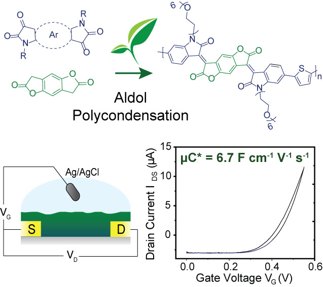
This research article highlights a new design strategy for n-type organic semiconductors combining sustainable synthesis with the high ionic/electronic conductivity needed for organic electrochemical transistors (OECT). The use of electron-deficient lactone building blocks allowed synthesis via Aldol polymerization, offering the advantage of avoiding toxic and environmentally harmful compounds, such as the organotin reagents commonly used in Stille polymerization. These conjugated polymers are an excellent choice for n-type OECTs, pushing towards a new generation of high-performing materials that are better for the environment.
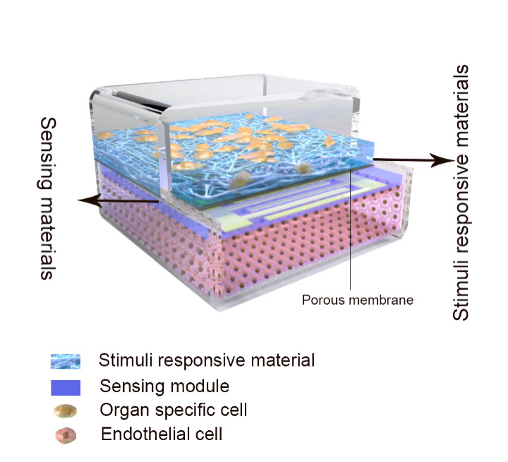
Advanced in vitro cell culture, microphysiological systems (MPSs), recapitulate features of human tissues and are increasingly being used for drug development and disease modeling. Still, they are commonly based on standard polymers with minimal real-time stimuli and read-out capacity. This review article describes how advanced materials and devices could enable a technology leap in reproducing in vivo-like functionality and real-time tissue monitoring.
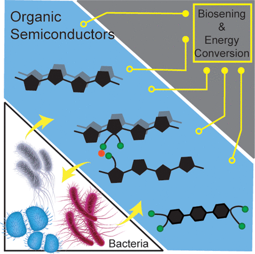
This review article summarizes the biological and electronic considerations when interfacing organic semiconductors with bacteria. From the biological perspective, it highlights the various mode employed by bacteria to communicate with the environment and with other bacteria. From the application perspective, it summarizes the characterization techniques and device geometries used to interface organic electronics with bacteria.
The article “Sorption of Neuropsychopharmaca in Microfluidic Materials for In Vitro Studies” was published in ACS Applied Materials & Interfaces today. The study highlights the impact of peristaltic pump tubing in sorption of hydrophobic compounds. mainly consisting of neuropsychopharmaca. The article further displays that the use of PDMS or other device construction methods OSTE+ or PC/PSA had a similar effect on the sorption, whereas the material of the tubing had a stronger dependence on sorption as compared to the device material. This signifies that the tubing and associated materials deserve similar attention as other device materials used for in-vitro studies.
Dr. Rohollah Nasiri joins the Herland group as our newest postdoctoral scholar. He received his PhD and MSc degrees in Mechanical Engineering from the Sharif University of Technology, Iran, in 2021 and 2014, respectively. His research focusses on designing organ-on-a-chip devices integrated with biosensors for disease modeling and drug screening applications.
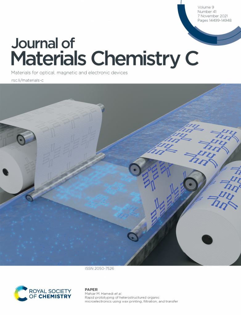
The article “Rapid prototyping of heterostructured organic microelectronics using wax printing, filtration, and transfer” was published in the Journal of Materials Chemistry C today. The research highlights rapid prototyping of various micropatterned organic electronic heterostructures of PEDOT:PSS using hydrogels filtered onto membranes containing hydrophobic wax patterns. The article also demonstrates the potential of this method for micro-supercapacitors, organic electronic transistors, and their use in cell culture to enable bioelectronics.
The article was also featured on the front cover of the journal.
The article “Continuous Monitoring Reveals Protective Effects of N-Acetylcysteine Amide on an Isogenic Microphysiological Model of the Neurovascular Unit” was published in Small today. The article reports a microphysiological blood-brain barrier model that captures the multicellular interactions of iPS-derived cells. The integrated electrical sensors, facilitated by PDMS-free fabrication, allow for real-time monitoring of how the barrier responds to oxidative stress and antioxidant prophylaxis. The sensor integrated hiBBB-on-chip displayed an immediate utility in the screening of drugs modulating the barrier by providing readout about the temporal pharmacodynamic profiles.
The article was also featured on the inside Back cover of the journal.
We have an open Postdoc (scholarship) position in the field of electrochemical biosensors. The post doc scholarship will focus on organic electrochemical transistors-based biosensors for the enzymatic sensing of small molecules. For more details and application, check the link below.
We have an open Postdoc (scholarship) position in the Wallenberg foundation funded project Organs-on-Chips for Translational Research in Brain Disease. This project will be focusing on inborn errors of metabolism in children, combining neural models with real-time sensing of neural function, barrier function and metabolic activity. Conventional cell culture, as well as microfluidic Organ-on-Chip methods will be applied. The post doc scholarship will focus on developing functional stem-cell derived cells for modelling inborn errors of metabolism combined with Brain-on-Chip systems. The postdoc will work closely together with engineers in the Herland lab and clinical researchers at Karolinska Hospital. More details can be found here.
Prof. Anna Herland was featured in an interview regarding research in the field of development of brain-on-chip models for testing new treatment methods and regimes on the KTH webpage. (Swedish only)
Erica Zeglio, our post-doc was featured in an interview with Vattenfall, a Swedish energy company where she gave a comment about an innovative technology for power generation using conducting bacterial nanowires.
Congratulations to our postdoc Erica Zeglio on being awarded a prestigious Marie Skłodowska-Curie Individual Fellowship by the European Commission for her project entitled “BioResORGEL: Bioresorpable Organic Electronic Devices”.
We have another open Postdoc (scholarship) position in the Wallenberg foundation funded project Organs-on-Chips for Translational Research in Brain Disease. The post doc scholarship will focus on developing and integrating sensors in Brain-on-Chip systems using conventional cell culture and microfluidic Organ-on-Chip methods. More details can be found here.
We have an open position for a Postdoctoral Scholarship in the Wallenberg funded project ‘Organs-on-Chips for Translational Research in Brain Disease’. More details can be found here.
Organs-on-Chips will be critical to reducing animal experiments in the future. There are manychallenges still to overcome toward their widespread use, one important challenge being the high cost and complexity of manufacturing current systems, both commercially or in an academic setting. In our work, we showcase a functional Barrier-on-Chip of the small intestine, fabricated using very simple and low-cost methods that practically anyone can implement in their lab, but that can also be adapted to large-scale industrial manufacture. We hope that this can make Barrier-on-Chip technology much more broadly accessible, particularly to researchers and prototypers in low-resource environments.
Comments from Prof. Anna Herland on the feature in ATLA